
Advanced surgical management for primary liver cancer, metastatic liver tumours, hilar cholangiocarcinoma, complex liver cysts, and localized infections, tailored around dynamic imaging, functional reserve metrics, and diagnostic staging.
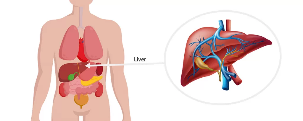
The liver is a massive, highly vascular organ responsible for filtration, protein synthesis, and metabolic regulation. Because it has the unique biological ability to regenerate, surgical interventions are focused on removing diseased segments while safeguarding enough healthy tissue to sustain critical body functions.
Liver Surgery Covers More Than One Condition
Hepatocellular Carcinoma (Primary Liver Cancer)
A primary malignancy arising from the functional liver cells (hepatocytes), often occurring on a background of chronic liver disease, fatty liver, or cirrhosis. Surgical removal requires careful balancing of tumor boundaries against the remaining functional capacity of the liver tissue.
Liver Metastases (Secondary Tumours)
Cancerous growths that have spread to the liver from an original site elsewhere in the digestive tract, most commonly colorectal cancer. Resecting these secondary lesions can be highly effective and is often closely coordinated alongside systemic chemotherapy cycles.
Hilar Cholangiocarcinoma
A complex malignancy developing in the major bile ducts right where they exit the liver (the hilum). Surgery often requires a combined approach—removing the affected liver lobe alongside the diseased bile ducts and reconstructing the fluid channel directly to the small intestine.
Benign Cystic Lesions & Abscesses
Non-cancerous fluid-filled sacs or complex infectious pockets (abscesses) within the liver tissue. While small cysts are left alone, large or complex cysts that cause pain, obstruction, or persistent internal infections require definitive unroofing, surgical drainage, or localized removal.
Liver Surgery in Ahmedabad: Procedures We Perform
Liver operations, globally termed hepatectomies, involve removing a precise architectural zone of the liver based on its internal blood vessel and bile duct anatomy.
Major Hepatectomy (Right or
Left Hemihepatectomy)
Removal of an entire anatomical half of the liver (either the right or left lobe) to achieve clear margins around large tumours, such as primary hepatocellular carcinoma or deep-seated hilar cholangiocarcinoma.
Before scheduling, advanced imaging ensures that the remaining side of the liver is fully healthy and large enough to expand and support the body’s metabolic needs post-operation.
Minor / Segmental Liver
Resection
Removal of one or more small, isolated anatomical segments of the liver rather than an entire lobe. This targeted approach is highly utilized for clearing accessible liver metastases, localized benign tumours, or superficial cystic lesions.
Because it preserves the maximum amount of normal tissue, it can often be performed via advanced laparoscopic or robotic access.
Localized Ablation (Microwave /
Radiofrequency Ablation)
A minimally invasive thermal technique where a specialized needle-like probe is guided directly into a liver tumor using ultrasound or CT imaging. High-frequency thermal energy is applied to destroy the cancerous cells on-site without removing the surrounding liver tissue.
This is an invaluable option for small tumours in patients whose overall liver health or medical fitness prevents a formal surgical resection.
Surgical Drainage of
Complex Abscesses
While standard liver abscesses respond well to antibiotics or simple needle aspiration, deep, multi-pocketed, or thick-walled abscesses that fail to resolve may require formal laparoscopic or open surgical clearing. The pus-filled sac is opened, thoroughly evacuated, and a dedicated drainage tube is placed to ensure the cavity collapses and heals from within.
Before Liver Surgery: What to Expect
Evaluating the liver’s underlying health is just as critical as mapping the tumor itself. The pre-operative phase follows a strict clinical pathway.
1. Functional Liver Evaluation
Blood panels, including liver function tests, viral hepatitis screenings, and coagulation profiles, are performed to assess how well the liver manufactures essential proteins and processes wastes.
2. High-Resolution Staging
Triple-phase CT scans of the abdomen or dedicated liver MRIs are utilized to track the exact relationship between the tumor and the major internal blood vessels (hepatic artery, portal vein, and hepatic veins).
3. Volumetric Assessment
For major resections, specialized software calculates the precise volume of the liver segment that will remain after surgery (the Future Liver Remnant). If this remaining segment is too small, alternative staged interventions are considered first.
4. Tumour Board Review
Malignant cases are structured within a multidisciplinary team format to decide if chemotherapy or localized down-staging is required before moving forward with a surgical plan.
About Dr Sourabh Damani
Practising as a Gastrointestinal and Laparoscopic Surgeon in Ahmedabad, with a focused interest in hepato-pancreato-biliary (HPB) interventions, structural liver resections, and multimodal management of abdominal malignancies.

Performs anatomical and non-anatomical liver resections, segmentectomies, and unroofing of complex cystic lesions.
Manages structural interventions for hepato-biliary tumours and coordinates treatment options for secondary liver metastases.
Facilitates clinical evaluations for advanced liver disease, structural complications of cirrhosis, and coordinates diagnostic pathways for transplant-eligible profiles.
Employs minimally invasive and ultrasound-guided approaches to maximize normal tissue preservation and support post-operative liver regeneration.
Liver Surgery in Ahmedabad: Frequently Asked Questions
How does the liver function after a large portion of it is surgically removed?
The liver possesses a unique capacity to regenerate. When a portion is removed, the remaining healthy liver cells begin to divide and grow rapidly. Within a few weeks to months, the liver can regenerate back to near its original volume and fully handle the body’s metabolic requirements, provided the remaining tissue was healthy before surgery.
What is the difference between a primary liver tumor and a liver metastasis?
A primary liver tumor (such as hepatocellular carcinoma) starts directly within the cells of the liver itself, usually linked to chronic liver issues or inflammation. A liver metastasis is a secondary growth that began as a cancer in another organ—like the colon or stomach—and shed cells that traveled through the bloodstream to settle in the liver. Their surgical and medical treatment protocols are quite distinct.
When is radiofrequency or microwave ablation chosen over standard liver surgery?
Thermal ablation is typically considered for patients who have small, localized liver tumours but cannot safely undergo a formal hepatectomy due to advanced age, poor underlying liver health (such as advanced cirrhosis), or other medical complications. It allows target clearance while completely avoiding the stress of major tissue removal.
What are the main signs that indicate a liver condition might require a surgical evaluation?
While many early liver issues are silent, advanced structural problems, large cysts, or expanding tumours often present with jaundice (yellowing of the eyes and skin), unexplained weight loss, persistent pain or a heavy sensation in the upper right side of the abdomen, or unexplained swelling in the legs and abdomen.
Please bring all recent high-resolution abdominal CT or MRI scan discs, triple-phase liver panels, tumor marker reports (such as AFP), and documentation of any past medical or oncological treatments. The initial consultation focuses on reviewing these parameters to verify surgical eligibility and establish a safe treatment sequence.